Intravenous Biomaterial Therapy: A Step-by-Step Guide to Internal Tissue Repair
Overview
Imagine repairing damaged heart muscle or brain tissue without surgery—simply by injecting a smart material into a vein. That’s the promise of a revolutionary biomaterial developed by scientists that travels through the bloodstream, homes in on injured areas, reduces inflammation, and kick-starts healing. Unlike earlier treatments that required direct injection into the heart, this therapy is delivered intravenously, allowing it to spread evenly and work rapidly. In animal studies, it successfully treated heart attack damage and showed potential for traumatic brain injury and pulmonary hypertension. This guide breaks down how this breakthrough works, what you need to know before applying it, and how to avoid common pitfalls.
Whether you’re a researcher, clinician, or simply fascinated by medical innovation, this tutorial will walk you through the entire process—from understanding the biomaterial’s design to administering it effectively.
Prerequisites
Before diving into the step-by-step instructions, it's important to have a basic grasp of the following concepts:
- Biomaterials: Knowledge of injectable hydrogels or self-assembling peptides that form nanofibers in the body.
- Inflammatory response: Understanding how inflammation plays a dual role in healing and scarring.
- Intravenous delivery: Familiarity with IV administration techniques and sterile protocols.
- Target conditions: Awareness of myocardial infarction, traumatic brain injury, and pulmonary hypertension pathology.
No prior experience with this specific therapy is required—the guide will cover everything from first principles.
Step-by-Step Instructions
Step 1: Prepare the Biomaterial Solution
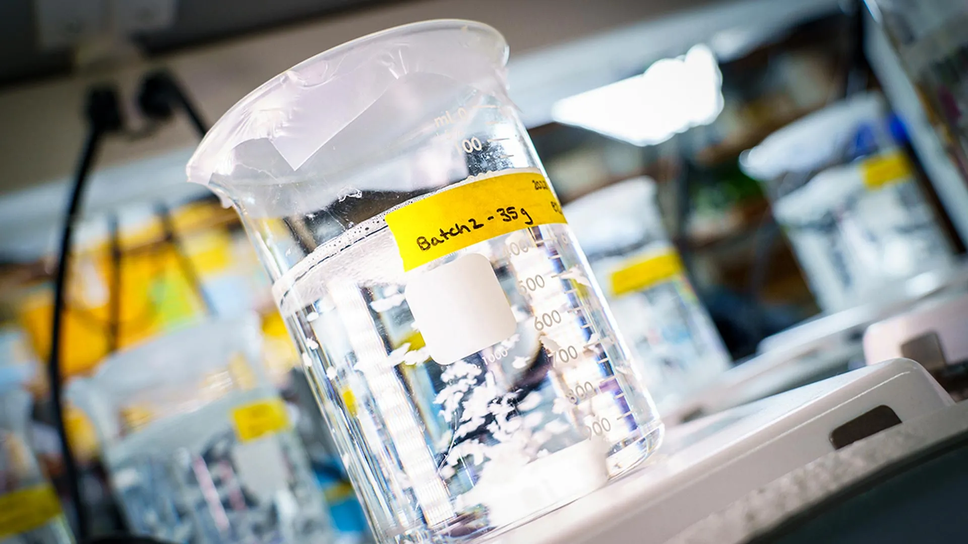
The biomaterial is a specially engineered peptide or polymer that self-assembles into nanofibers when exposed to physiological conditions. In the lab, the lyophilized powder is reconstituted with sterile saline or buffer to a precise concentration (typically 50–100 mg/mL, depending on the formulation). The solution must be filtered through a 0.22 µm sterilizing filter to remove any aggregates or contaminants.
Important: The material should be prepared fresh and used within 30 minutes to avoid premature self-assembly.
Step 2: Administer Intravenously
The patient is positioned comfortably, and a standard IV catheter is placed in a peripheral vein—most often in the arm or hand. The prepared solution is slowly injected using a syringe pump at a rate of 2–5 mL/minute to prevent rapid bolus delivery. The total volume depends on the condition treated: for acute myocardial infarction, a dose of 0.1 mL/kg body weight is used in animal models; clinical trials may vary.
After injection, flush the line with 5 mL of saline to ensure the entire dose enters circulation.
Step 3: Circulation and Targeting
Once in the bloodstream, the biomaterial circulates as soluble monomers. Its surface is decorated with bioactive peptides that bind specifically to markers of injury, such as exposed collagen or fibrin from damaged blood vessels. This “homing” mechanism ensures the material accumulates only in injured tissues, not healthy ones.
Within minutes, the monomers are drawn to the inflamed site by chemotactic gradients and start assembling into nanofiber networks.
Step 4: Self-Assembly and Binding at the Injury Site
At the damaged tissue, local pH, ionic strength, or enzymatic activity triggers rapid self-assembly. The nanofibers form a mesh that physically supports the tissue, similar to a scaffold. Additionally, the fibers present ligands that bind to integrins on cells, promoting cell adhesion and migration.
This step is critical: the biomaterial must maintain its structure for at least 24–48 hours to allow the body's own repair processes to initiate.
Step 5: Modulate Inflammation
The biomaterial reduces inflammation by two mechanisms:
- Scavenging free radicals: Certain chemical groups within the material neutralize reactive oxygen species (ROS) produced by immune cells, preventing further tissue damage.
- Signaling to immune cells: The material releases anti-inflammatory cytokines (e.g., IL-10) or sequesters pro-inflammatory ones, shifting macrophages from M1 (pro-inflammatory) to M2 (pro-healing) phenotype.
This dampens the inflammatory cascade and prevents excessive scarring.
Step 6: Stimulate Tissue Regeneration
As inflammation subsides, the biomaterial provides a scaffold for stem or progenitor cells to repopulate the area. Growth factors (like VEGF, FGF) are either incorporated into the material or released by attracted cells. Over the course of days to weeks, new blood vessels form and functional tissue regenerates—whether heart muscle, neurons, or lung tissue.
Eventually, the biomaterial degrades into harmless byproducts (e.g., amino acids) and is cleared by the kidneys.
Common Mistakes
Mistake 1: Incorrect Solution Concentration
If the biomaterial is too dilute, it won’t form stable fibers at the injury site—it may simply wash out. If too concentrated, it can clog the IV line or cause premature aggregation in circulation. Always verify the exact concentration recommended for the specific formulation.
Mistake 2: Rapid Injection
Injecting too quickly can trigger a systemic inflammatory response or cause the material to aggregate in the lungs. Use a slow, steady rate (≤5 mL/min). Monitor for any signs of difficulty breathing or hypotension.
Mistake 3: Ignoring Patient Selection
This therapy is not suitable for all conditions. For example, patients with severe kidney impairment may not clear the degradation products efficiently. Similarly, those with active infections could experience exacerbated inflammation. Screening for organ function and infection status is essential.
Mistake 4: Overlooking Timing After Injury
The biomaterial works best when administered within 1–6 hours after tissue damage (e.g., heart attack or brain injury). Delayed injection may find the tissue already irreversibly scarred or the biological window closed. Adhere to the therapeutic time window defined in the protocol.
Summary
The intravenous biomaterial therapy represents a paradigm shift in regenerative medicine—no surgery needed, precise targeting, and dual action of inflammation reduction and tissue repair. This guide walked you through the six steps: preparation, administration, circulation, self-assembly, immune modulation, and regeneration. By avoiding common mistakes like incorrect concentration, rapid injection, poor patient selection, and wrong timing, you can maximize therapeutic success. While still in preclinical stages, the promise for treating heart attacks, brain injuries, and pulmonary hypertension is immense.
Related Articles
- 5 Key Insights into the FDA's New Acting Vaccine and Biologics Director
- The Arginine Approach: A Step-by-Step Guide to Potentially Reducing Alzheimer’s Damage with a Common Amino Acid
- FDA Investigates: Cancer-Causing PFAS Chemicals Detected in Multiple Brands of Infant Formula
- Harnessing Blood-Based DNA Markers to Monitor Arsenic Exposure and Predict Health Risks
- Federal Court Restricts Mail Delivery of Abortion Pill: Key Questions Answered
- Understanding PFAS in Baby Formula: Key Questions Answered
- 6 Unseen Realities Faced by Older Homeless Women
- Decoding the Mechanism: How a Common Cold Virus Blocks Cancer Metastasis to the Lungs